Advanced Diagnostic Testing
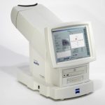
Optical Coherence Tomography (OCT) is a non-invasive diagnostic instrument used for imaging the retina. It is the technology for the future because it can enhance patient care. It has the ability to detect problems in the eye prior to any symptoms being present in the patient. With an OCT, doctors are able to see a cross section or 3D image of the retina and detect the early onset of a variety of eye conditions and eye diseases such as macular degeneration, glaucoma and diabetic retinopathy (the top three diseases known to cause blindness).
The OCT allows for detection of other diseases such as macular holes, hypertensive retinopathy and even optic nerve damage. Using an OCT allows for early treatment in patients and dramatically improves the success of these treatments, especially in diseases such as wet macular degeneration – where the eye disease progresses rapidly.
The OCT has become the standard of care for the assessment and treatment of most retinal diseases, it is similar to a CT scan which is used to image internal organs inside the body. The OCT uses an array of light to rapidly scan the eye. These scans are interpreted and the OCT then presents an image of the tissue layers within the retina. These layers can be differentiated and their thickness measured. By comparing the thickness of the layers measured by the OCT scan against the normal thickness of healthy retinal layers, eye doctors can determine which retinal disease or eye condition exists in the eye, even before the patient is aware of any problems

Visual Fields Testing
Humphery Vision FieldsA visual field test is a method of measuring an individual’s central and peripheral (side) vision. Visual field testing is most frequently used to detect any signs of glaucoma damage to the optic nerve. In addition, visual field tests are useful for detection of central or peripheral retinal disease, eyelid conditions such as ptosis or drooping of the eyelid, optic nerve disease, and diseases affecting thePerimeter.
visual pathways within the brain. Many eye and brain disorders can cause peripheral vision problems and visual field abnormalities. Brain abnormalities such as those caused by strokes or tumors can affect the visual field. In fact, the location of the stroke or tumor in the brain can frequently be determined by the size, shape and site of the visual field defect.
To do the test, you sit and look inside a bowl-shaped instrument called a perimeter. While you stare at the center of the bowl, lights flash. You press a button each time you see a flash. A computer records the spot and at the end of the test, a printout shows if there are areas of your vision where you did not see the flashes of light. These are areas of vision loss. If you are diagnosed with a particular disorder or disease such as glaucoma, visual field testing will become a routine part of your treatment. After a baseline has been established, visual field testing is repeated every 6 to 12 months to monitor for change. Visual field tests plays a critical role in helping the doctor follow your condition
Digital Retinal Photography
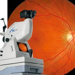
Have you ever seen the inside of your eye? See what the doctor sees when he looks into your eyes! We feel that it is important that our patients understand what we are looking for, and what we see when the doctor looks inside your eyes.
Many eye problems can develop without warning and progress with no symptoms. Early on, you might not notice any change in your vision. However, diseases such as macular degeneration, glaucoma, retinal tears or detachments, central retinal vein or artery occlusion, optic strokes, as well as other health problems such as diabetes and high blood pressure, can be detected with a through retinal examination utilizing a digital retinal camera.
Digital retinal photography consists of a digital camera system that takes a photograph of your retina. Here is a summary of the benefits of Digital Retinal Photography:
- A scan to confirm a healthy eye or detect the presence of disease.
- An overview or map of the retina, giving your eye doctor a more detailed view than he can achieve by other means.
- The opportunity for you to view and discuss the images of your eye with your doctor at the time of your examination.
There is no need to wait for results, our digital camera gives us the images within seconds for review. By looking at digital photos from previous years, your doctor can look for changes over time and determine if more aggressive treatments are necessary.
 We recommend that all our patients receive this test. It is especially important for patients with a history of high blood pressure, diabetes, retinal diseases, flashes of light, floaters, headaches and those with a strong glass prescription.